Lower Limb Venous Duplex
Assessing for Varicose Veins
- Map out normal & abnormal venous pathways
- Evaluate blood cloths
- Safe & painless ultrasound scan
Duplex ultrasound scanning is widely regarded as a “gold-standard” procedure, providing medical professionals with detailed imagery to manage the concern.
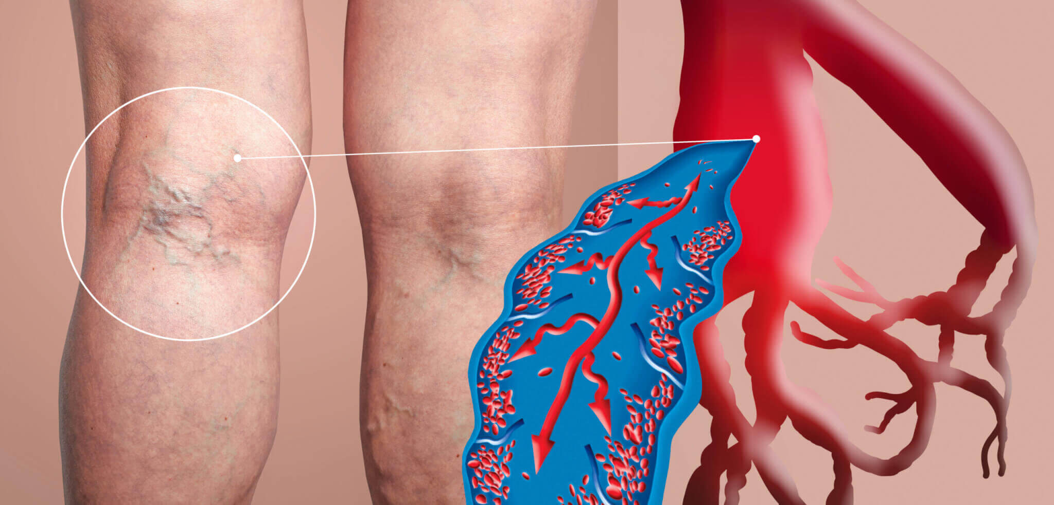
There are many veins which return blood from the legs to the heart. The valves within these veins are responsible for controlling and directing the flow of blood in the right direction. In the case of varicose vein disease these valves fail, allowing blood to be drawn back by gravity. The result – bulging of the veins on the surface of the legs. The long term consequence of this occurrence can include aching and painful legs, skin changes, swelling or ulceration.
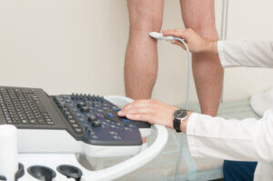
A venous duplex ultrasound scan is a safe and non-invasive diagnostic test administered to assess varicose veins and other circulatory disorders. The screening includes the use of an ultrasound “doppler” to assess blood flow. The doppler provides precise information on the vascular anatomy as well as determining the speed and direction of blood flow. It is able to provide detailed examination of the great and small saphenous veins, as well as the deep veins in which clots (DVT) sometimes occur.
Venous duplex ultrasound scans provide detailed information which forms a map of patient veins. This is essential when planning the correct treatment, thereby ensuring the optimum result and minimising the chances of future vein recurrence.
This type of ultrasound scan is usually performed whilst patients are standing.


Ⓒ Bodyvie Limited 1999 - 2024 All rights reserved. All trademarks acknowledged.
Company number 3849113
